Mohs Micrographic Surgery
Introduction
Mohs micrographic surgery, a surgical treatment for skin cancers that offers a very high rate of cure, was developed by Dr. Frederick E. Mohs, M.D. This technique of Mohs surgery is time-consuming and requires highly specialized training and personnel. You were referred to our practice because Mohs micrographic surgery has been proven to be a highly successful form of treatment for your type of skin cancer. I completed a fellowship in Mohs micrographic surgery following my dermatology residency.
This website was designed to give you information about skin cancer, as well as discuss what you can expect on the day of your surgery and during the period after surgery. We have also included a section that outlines preventative measures you can take to decrease the possibility of developing skin cancers in the future. Please read this section in its entirety before surgery. If you have any questions, please call Dr. Dozier's office at (512) 527-9020 and ask to speak to a member of the medical staff.
What You Should Know About Mohs Surgery
Mohs micrographic surgery is a specialized technique for the total removal of skin cancers. The procedure is done in several steps (see diagram below): 1) A local anesthetic (numbing medicine) is injected around the skin cancer. 2) The visible cancer is removed by curettage (scraping). 3) The area of the cancer is removed in a specific fashion so that the entire undersurface and skin edges can be examined microscopically. 4) The tissue is dyed and a map is drawn so that tumor found microscopically can be located on the patient. The tissue is then processed into microscopic slides. 5) The surgeon then examines the slides under the microscope for cancer cells. If any cancer is found, it will be marked on the map to guide the surgeon in removing additional tissue. 6) Subsequent layers of tissue are removed in a similar manner until no more cancer cells are found. In almost all cases, the surgery is done on an out-patient basis.
The Mohs Surgery Process
Step 1
The roots of a skin cancer may extend beyond the visible portion of the tumor. If these roots are not removed, the cancer will recur.
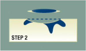
Step 2
The visible tumor is surgically removed.
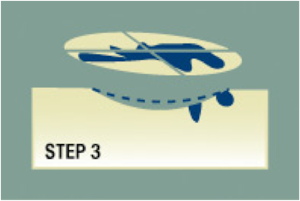
Step 3
A layer of skin is removed and divided into sections. The ACMS surgeon then color codes each of these sections with dyes and makes reference marks on the skin to show the source of these sections. A map of the surgical site is then drawn.
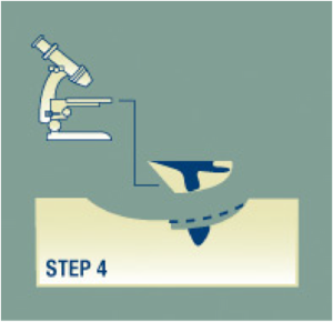
Step 4
The undersurface and edges of each section are microscopically examined for evidence of remaining cancer.
Step 5
If cancer cells are found under the microscope, the ACMS surgeon marks their location onto the "map" and returns to the patient to remove another layer of skin - but only from precisely where the cancer cells remain.
Step 6
The removal process stops when there is no longer any evidence of cancer remaining in the surgical site. Because Mohs surgey removes only tissue containing cancer, it ensures that the maximum amount of healthy tissue is kept intact.
How To Prepare For Your Surgery
Try to get a good night’s sleep.
Wash your hair either the night before or the morning of surgery.
Eat a normal breakfast.
Wear a shirt that buttons down the front; avoid “pullover” style clothing.
Do not wear makeup or jewelry if surgery is to be performed on your face.
Take any medicine as you normally would (this includes aspirin and other blood thinners) unless specifically instructed not to by Dr. Dozier; if you have been prescribed preoperative antibiotics, take them as directed and bring them with you on the day of surgery.
Bring a friend or relative to keep you company during the day; a good book or magazine would also be helpful. Surgery done on the face, particularly around the eyes or nose, can involve bandages or temporary muscle paralysis that may affect your vision. It is advisable to arrange to have someone bring you to and pick you up from your Mohs surgery appointment unless Dr. Dozier tells you otherwise.
Certain things can cause increased problems with bleeding during and after surgery. To decrease the risk of excess bleeding, follow the instructions below carefully:
DO NOT take aspiring or aspirin-containing products (such as Bufferin, Anacin, Ecotrin or Alka Seltzer) for two weeks before your surgery unless aspirin is part of the medication regimen prescribed or recommended by your doctor.
DO NOT take ibuprofen or naproxen sodium products (such as Advil, Motrin, or Aleve) for one week prior to surgery; pain relievers such as Tylenol or acetaminophen do not cause problems with bleeding and may be taken during the two weeks preceding surgery.
DO NOT take vitamin E or herbal supplements one week before surgery.
DO NOT drink alcohol (including wine and beer) for 24 hours before surgery and 48 hours after.
DO NOT plan to engage in any heavy lifting or exercise for at least 48 hours after your surgery; it is best to resume your normal exercise only after your stitches have been removed.
What To Expect On The Day Of Surgery
Shortly after your arrival, you will be taken to one of the treatment rooms, where I will again review with you the risks and benefits of Mohs micrographic surgery and have you sign a written consent. The area of the skin cancer will then be cleaned and a local anesthetic will be used to numb the area. I will remove the cancer and a thin layer of skin surrounding the cancer. After this has been done, any bleeding will be stopped with an electric instrument called a cautery. You will then be bandaged and be able to relax in the waiting room while the tissue that was removed is being processed. It usually takes 20 to 30 minutes for the layer of tissue to be removed and the bleeding to stop. However, it takes about one to one and a half hours for the tissue to be prepared into microscopic slides for examination. During this time you may chat with the person accompanying you, read a book or watch television.
If examination of the microscopic slides reveals that your tissue still contains tumor cells, the procedure will be repeated. Further tissue is removed only from the areas where tumor cells were found. The goal is to remove all of the skin cancer while preserving the greatest amount of healthy tissue. However, skin cancers can grow deeply and develop roots extending beyond the area you can actually see. As a result, the final size of the surgical incision will be determined by the extent of the tumor. The average number of surgical sessions required is two to three. However, you may require more before your skin cancer is completely removed. Fortunately, this can usually be done in the course of a single day. When surgery is completed, a decision will be made as to the way to manage your wound.
What To Expect After Your Surgery
Pain:
Most patients do not have severe pain, but may experience slight discomfort. If this occurs, we suggest you take two tablets of Extra-Strength Tylenol every four hours. Avoid compounds containing aspirin, ibuprofen and naproxen sodium as you did before surgery. These products may cause bleeding. If this does not control the pain, take the pain medicine I have prescribed for you.
Bleeding:
Occasionally, bleeding follows surgery. If this happens, do not become alarmed. Lie down and place steady, firm pressure over the wound as close as possible to the bleeding area. Apply the pressure continuously for 20 minutes (timed). Do not lift the bandage to check on the bleeding. If the bleeding persists after 20 minutes of steady pressure, apply pressure for an additional 20 minutes. If the bleeding still continues contact me by calling (512)527-9020.
Swelling and bruising:
Swelling and bruising are very common after surgery, particularly when it is performed near the eye. All wounds swell a little. Usually this is not a problem, and the swelling and bruising decrease as the wound heals. The amount of swelling that occurs after surgery can be decreased by applying ice packs (such as a bag of frozen peas) to the area of surgery for periods of 20 minutes on and off the 24 hours after surgery.
Drainage:
All wounds drain to some extent during the first week or two. This is why frequent dressing changes are necessary.
Infection:
Infection of the wound is unusual. However, if you see thick, foul smelling fluid coming from the wound, call our office. An antibiotic may be necessary.
Redness:
All wounds will develop a halo of redness that disappears gradually. If the area becomes extremely red and itchy, you may be allergic to the antibiotic ointment or the tape. Call our office if this develops.
Scarring:
Most surgeries leave a scar. However, your scar will improve and become less noticeable as time passes.
Stitches and skin grafts:
If we close the wound with sutures (stitches) or place a skin graft, you should keep the area clean, bandaged as directed and dry until the next clinic visit. If you see foul-smelling fluid coming from the wound, call our office immediately for instructions on how to contact me. An infection may be developing and an antibiotic may be necessary.
What To Expect After The Wound Has Healed
You may experience some tightness, or drawing, as the wound heals. This is normal and usually lessens with time. Patients also commonly experience some itching after their wounds have healed. This is often relieved by rubbing a small amount of plain petroleum jelly on the scar. Frequently, tumors involve nerves, and it may take up to a year, or even two years, before normal feeling returns to an area. Sometimes the area remains numb permanently. Only time will tell. The scar tissue that grows over the wound contains many more blood vessels than the surrounding skin. This results in a red scar that may be sensitive to temperature changes. This sensitivity improves with time and the redness usually fades. If the redness persists and extra blood vessels appear at the margin of the scar, they can be decreased with VBeam laser treatment that can be performed anytime after the surgery. Rarely, scar irregularities develop that can be treated with regional Dermabrasion that is optimally performed six to eight weeks after the surgery. Otherwise, most scar revisions should be delayed for a period of six to twelve months as the scar continues to improve during this period of time.
What You Should Know About Skin Cancer
How skin cancer forms: Cancer of the skin, like other cancers, is a disease of the cells. Cells are tiny structures that make up all parts of the body. Although they differ in shape and function in various organs, all cells reproduce themselves by dividing. Normal growth and repair of tissue takes place in this orderly fashion. When cell division is not orderly and controlled, abnormal growth occurs. Masses of tissue called tumors build up. Tumors can be benign or malignant. A malignant tumor is a cancer. Benign tumors do not spread. But cancerous or malignant tumors invade and destroy surrounding normal tissue as they grow. Occasionally, cancer cells may break away from the tumor and spread (metastasize) through either the blood or lymphatic vessels to distant parts of the body, where they form additional tumors.
Types of skin cancer: The three most common forms of skin cancer are: basal cell carcinoma, squamous cell carcinoma and malignant melanoma. Most cases of skin cancer are either basal cell carcinoma or squamous cell carcinoma. Squamous cells make up most of the surface layer of the skin. Basal cells occur in smaller number and are located in the lowest part of the skin. Of the two, basal cell carcinoma occurs more frequently and grows more slowly. It very rarely spreads through the blood or lymphatic systems to distant parts of the body. Squamous cell carcinoma is the second most common skin cancer. It is more serious, since it has the ability to spread internally to nearby lymph nodes and to other parts of the body. Malignant melanoma may be life-threatening if not treated early. It usually appears as a brownish-black spot or bump in the skin that enlarges and sometimes bleeds. Sometimes melanomas arise in moles that have been present for years. Malignant melanoma is rarely treated with Mohs surgery.
Causes of skin cancer: Exposure to the sun appears to be the leading cause of skin cancer, which commonly develops on the face, neck and arms, the most sun-exposed areas of the body. Fair-skinned people develop skin cancer more frequently than dark-skinned people. Skin cancer also tends to be hereditary and occurs very frequently in certain ethnic groups, particularly those with fair skin. Other causes of skin cancer include x-ray exposure, chronic trauma and certain chemicals.
Signs of skin cancer: Although most skin growths are benign, any new growth on the skin or a sore that does not heal should be brought to your dermatologist’s or primary care doctor’s attention. Skin cancer has many different appearances. It may begin as a small, waxy lump that eventually bleeds and crusts; as a dry, scaly, red patch; or in several other ways. Although it may begin very small, skin cancer can grow to become very large. Skin cancer sometimes forms from a non-cancerous skin condition called actinic keratosis. These are red, rough patches of skin that develop as a result of sun damage. They are commonly found on the face, neck, or hands. If the doctor thinks that a skin growth may be cancerous, a biopsy is performed. The whole area or a sample of the area is removed surgically and sent for examination under the microscope. The biopsy is used to confirm or rule out the diagnosis of cancer and to determine the type of any cancer found.
Treatment of skin cancer: Several effective methods are used to treat skin cancer. Which treatment method is chosen depends upon factors such as the location of the cancer, its size and previous therapies. The treatment methods include surgical removal, or excision, with suturing (stitching); curettage and electrodessication, which is scraping and burning with an electric needle; radiotherapy (X-ray treatments); cryosurgery (freezing); immunomodulators; photodynamic therapy; and Mohs micrographic surgery. Many of these treatments have high cure rates, but Mohs micrographic surgery uniformly produces the highest success rate, especially for the most difficult tumors.
Defense Against Future Skin Cancer
The most important thing you can do to lessen the possibility of developing future skin cancers is to protect your skin from further sun damage. Try to avoid sum exposure during the hours of 10 a.m. to 4 p.m. For unavoidable sun exposure, take protective measures by daily application of sunscreens to all exposed skin, including the tops of the ears. You also may wish to obtain further protection by wearing a broad-brimmed hat and appropriate protective clothing. Sunscreens can be purchased in your pharmacy without a prescription. These are colorless creams or lotions that absorb or block the damaging rays of the sun that cause sunburn and other skin damage. Apply the sunscreen 15 minutes before you go outdoors. It may wash off with water or heavy perspiration, so be sure to reapply sunscreen after swimming or exercise, and use a sunscreen labeled waterproof. There are a number of excellent sunscreens available. Whatever sunscreen you choose, it should have a sun protection factor (SPF) of at least 15 and be broad spectrum (protecting against ultraviolet A and B).
Follow-Up Visits
Once your surgery is over and your sutures have been removed, periodic follow-up exams by your referring physician are important for at least five years. These visits can help with early detection and treatment of any new skin cancers that may develop as well as the rare recurrence of the skin cancer that was treated. As a person who has had a skin cancer, you are at risk of developing others in the years ahead. You should be seen at least once every year by your physician. If you notice any suspicious areas on your skin between visits, it is best to check with your physician to see if a biopsy is needed.
For further information regarding Mohs Micrographic Surgery you can visit The American College of Mohs Surgery web site.